Ultrasound
About This Procedure
During an ultrasound, the patient remains on an exam table and is positioned appropriately for the exam. A technologist applies a warm gel to the skin in order to transmit sound waves and a hand-held device is placed in contact with the gelled skin and maneuvered to complete the study. The exam is typically completed within 30 - 45 minutes, however, there are more involved procedures (more than one technologist, nurse and/or physician present, etc.) that may take up to 1 - 1.5 hours.
Prior to an ultrasound, a patient may be required to drink water ahead of time, in order to fill the bladder. Other preparations may include fasting or advance bowel preparation.
Common ultrasound uses
Ultrasound imaging, also called ultrasound scanning or sonography, is a noninvasive diagnostic tool which involves the use of high-frequency sound waves to produce pictures from inside of the body. Ultrasound images are captured in real-time, making them extremely effective in showing the structure and movement of many of the body's internal organs, as well as blood flowing through blood vessels.
Typically performed on an outpatient basis, Franciscan Health uses ultrasound to help physicians evaluate symptoms such as pain, swelling and infection. Ultrasound can also be used to guide procedures such as needle biopsies and to take picture of the breasts in helping to detect breast cancer.
Ultrasound is also used to monitor a fetus while still inside the mother's womb. Obstetrical ultrasounds often include pictures or even video images of the baby or babies.
Related Locations

Imaging +1 more
Franciscan Health Crown Point Diagnostic Imaging Center
2411 LaPorte Avenue - Valparaiso, IN 46383
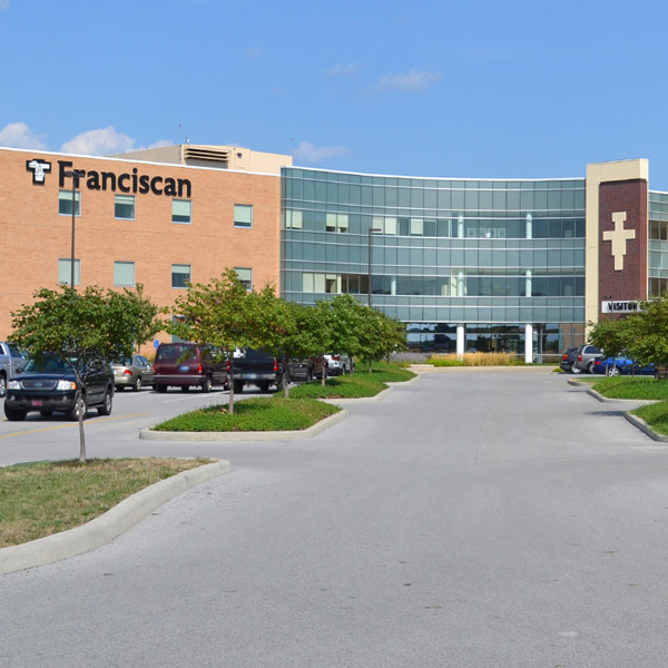
Imaging +1 more
Franciscan Health Imaging Lafayette
1701 S. Creasy Lane - Lafayette, IN 47905

Imaging +1 more
Franciscan Health Diagnostic Imaging Center Michigan City
3500 Franciscan Way - Michigan City, IN 46360