Endobronchial Ultrasound (EBUS)
About This Procedure
Any tools that we can use that help us detect lung disorders early, are powerful tools in the fight against cancer. Endobronchial ultrasound is one of those powerful tools. Endobronchial ultrasound (EBUS) is a more accurate and less invasive way of assessing a patient's lymph nodes to diagnose lung cancer, infections or other diseases. While performing an endobronchial ultrasound, doctors have real-time imaging of the surface of the airways, blood vessels, lungs and lymph nodes. Endobronchial ultrasound offers an alternative to surgery and has proven to be a highly effective tool in the treatment of lung cancer.
What happens during an endobronchial ultrasound?
An ultrasound guided endoscope or bronchoscope is passed through the mouth into the trachea. No incisions are needed. The airway and surrounding structures and lymph nodes can be viewed and sampled. The tests are highly accurate and often complement imaging diagnosis procedures. Performed together, the tests are more sensitive than either alone, and may become the next gold standard in lung cancer staging, the process of determining how much cancer is in the body and where it is located.
EBUS is typically performed as an outpatient procedure under moderate sedation and is often performed at the same time as electromagnetic navigational bronchoscopy.
Health Risk Assessment
What's your score? Take our LungCancerAware assessment, and in just minutes, receive a free, confidential analysis of your current risk factors for developing lung cancer.
Related Providers

Rebecca E Rizo, NP
Nurse Practitioner +1 more
Accepting New Patients
Offers Virtual Visits
Franciscan Physician Network

Related Locations
Hospital
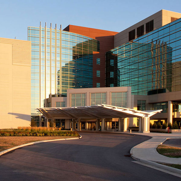
Franciscan Health Indianapolis
8111 South Emerson Avenue - Indianapolis, IN 46237
Hospital
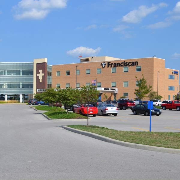
Franciscan Health Lafayette
1701 South Creasy Lane - Lafayette, IN 47905
Specialty Care +1 more
Franciscan Health Interventional Pulmonology Clinic Indianapolis
8111 South Emerson Avenue - Indianapolis, IN 46237